
Erler-Zimmer GmbH & Co. KG meningioma 3D model:
- Model code: MP2004
- Clinical history
- A 68-year-old woman presented with new-onset seizures and was diagnosed with epilepsy.
- A repeated history revealed gradual changes in the patient's personality.
- She later died of a myocardial infarction a few months later.
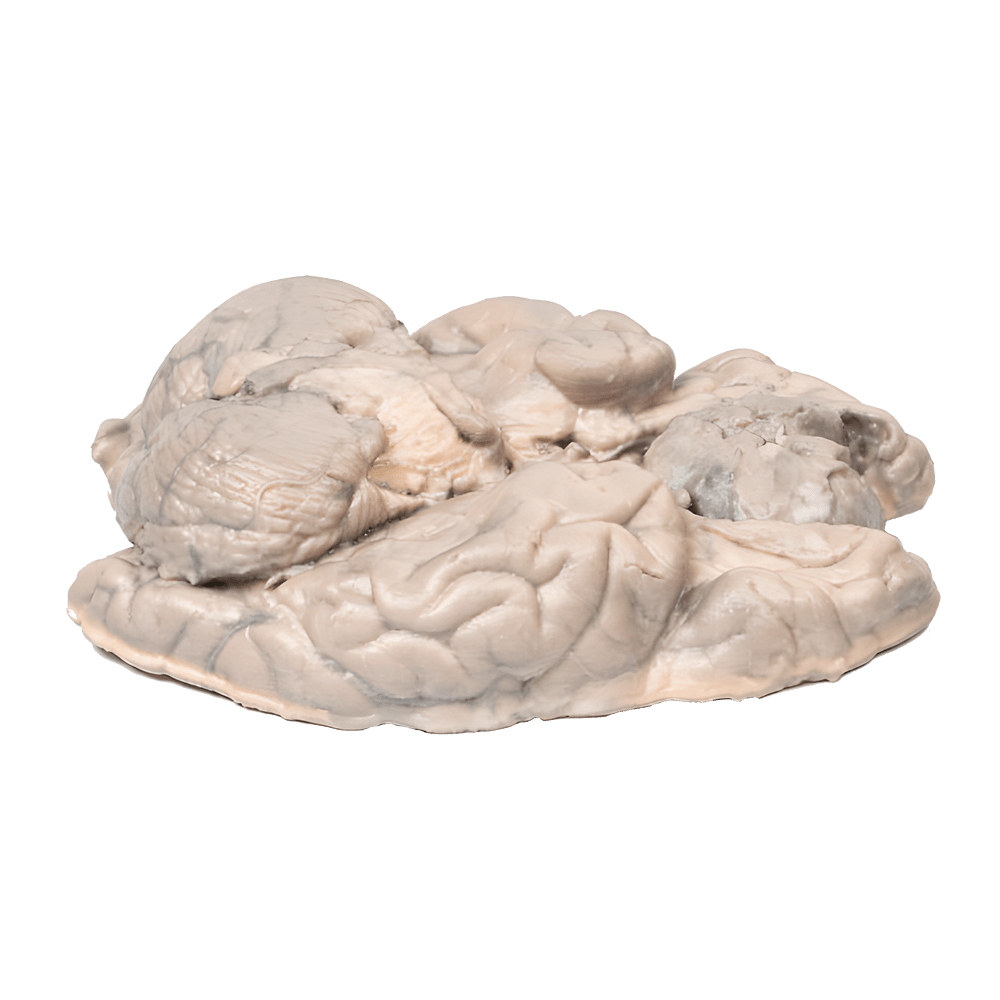
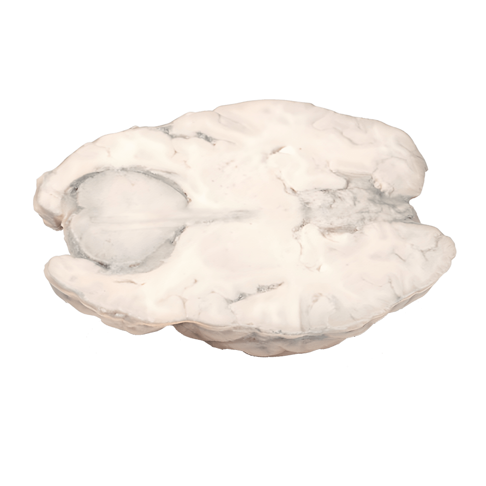
- Pathology
- This brain sample is horizontally sliced.
- A well-defined 6 cm tumor is seen between the two anterior ventricles.
- The tumor compresses the anterior ventricles.
- It has a reddish cut surface with a few yellow areas, indicating necrosis.
- It was attached to the dura mater anteriorly.
- This is an example of a meningioma.
- Read more
- Meningiomas are often considered the most common tumors of the central nervous system (CNS); however, they actually arise from the meninges (dura, arachnoid, and pia), which are not strictly speaking part of the CNS per se.
- They arise from arachnoid cells closely associated with the dura mater; therefore, these tumors may involve the dura mater or the dural arches (falx cerebri and tentorium cerebelli).
- Meningiomas are mostly slow-growing benign tumors.
- Symptoms depend on the location of the tumor and its growth rate.
- Symptoms include seizures, personality changes, changes in vision, hearing, or smell, and symptoms related to increased intracranial pressure.
- Meningiomas are often asymptomatic.
- Treatment includes observation, surgery, or radiation, depending on the clinical context and tumor morphology.
Meningioma 3D models – Erler-Zimmer Anatomy Group
More information: on the manufacturer's website or send us an inquiry!


